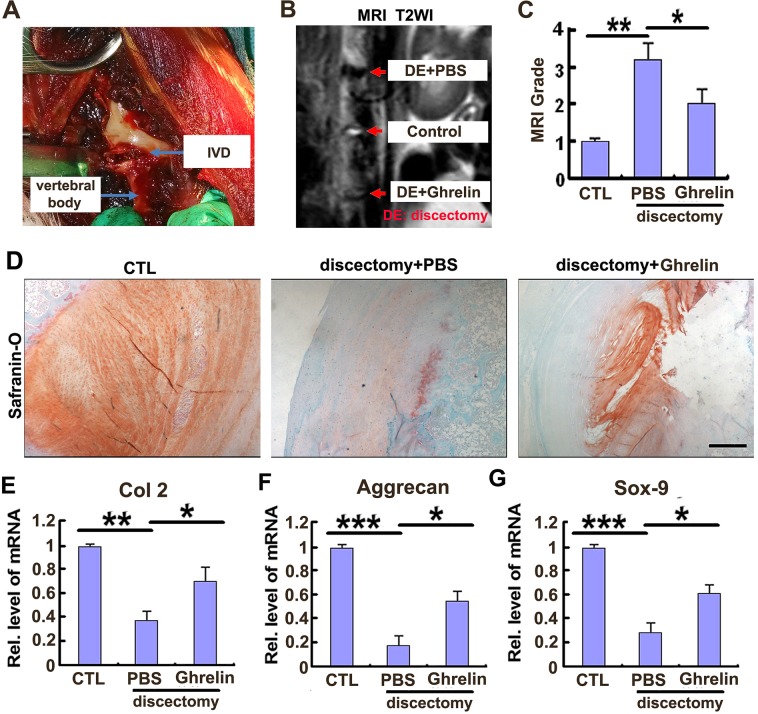
Figure 4. Ghrelin protects against destruction of NP tissue in a rabbit IVD degeneration model.
(A) The representative image during surgery. (B) Signal of IVD destruction was displayed in W2 weighted image in the rabbit model, while ghrelin treatment markedly improved the IVD structure, as assayed by MRI. 8 weeks after surgery, rabbits were anesthetized and MRI was performed in each group (N=7 per group). (C) Ghrelin reduced the MRI grade in IVD discectomy model, as indicated by MRI grading system. (D) Ghrelin treatment maintained extracellular matrix in NP tissue. IVD samples were isolated from each experimental group, and Safranin-O staining was performed. (E-G) Ghrelin treatment attenuated the reduction of Col 2, aggrecan as well as Sox-9 in NP tissue, as assayed by real time PCR. NP tissue was collected and real time PCR was performed. The values are the mean±SD. *p < 0.05, ** p < 0.01 and *** p < 0.005 vs. Control group. Each experiment was repeated for 3 times. Scale bar=250μm.

