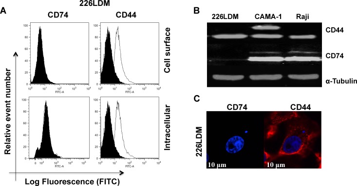
Figure 4. The expression of CD74 and CD44 receptors in immortalized normal breast luminal cells (226LDM).
(A) Cell-surface and intracellular expression of CD74 and CD44 was acquired by flow cytometry using By2 (anti-CD74) and 156-3C11 (anti-CD44). Black-filled histograms represent the 226LDM cells stained with indicated antibody. Empty histograms show the isotype as negative controls. (B) Total protein of CD74 and CD44 was detected by Western blotting, and α-tubulin was used as a loading control. (C) Confocal images of CD74 and CD44 in 226LDM cells stained intracellularly: CD74 was labelled with Alexa Fluor 488 (green) and CD44 with Alexa Fluor 555 (red). Figures depict representative samples from duplicate experiments. Scale bar 10 μm.

