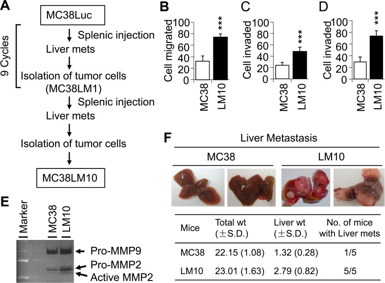
Figure 1. LM10 cells display more aggressive characteristics than parental MC38 cells.
(A) Schematic representation of experimental flow chart, showing the generation of LM10 cells after 10 cycles of splenic injection. (B) Migration assay was performed to assess the aggressive characteristics of LM10 cells. Data are presented as mean ±SD from three independent wells. ***p<0.001 when compared with control. (C and D) Cell invasion assay through collagen (C) or matrigel (D) was performed as described in Materials and Methods. Data are presented as the mean ±SD from three independent wells. ***p<0.001. (E) Gelatinase zymographic analyses of MMP-2 and MMP-9 activity were performed using parental MC38 and LM10 cells. Both MMP-2 and MMP-9 activity was increased in LM10 cells compared to parental MC38 cells. (F) MC38 and LM10 cells were injected into spleens of C57BL/6 mice (5 mice in each group). Liver metastasis was assessed after four weeks of splenic injection.

