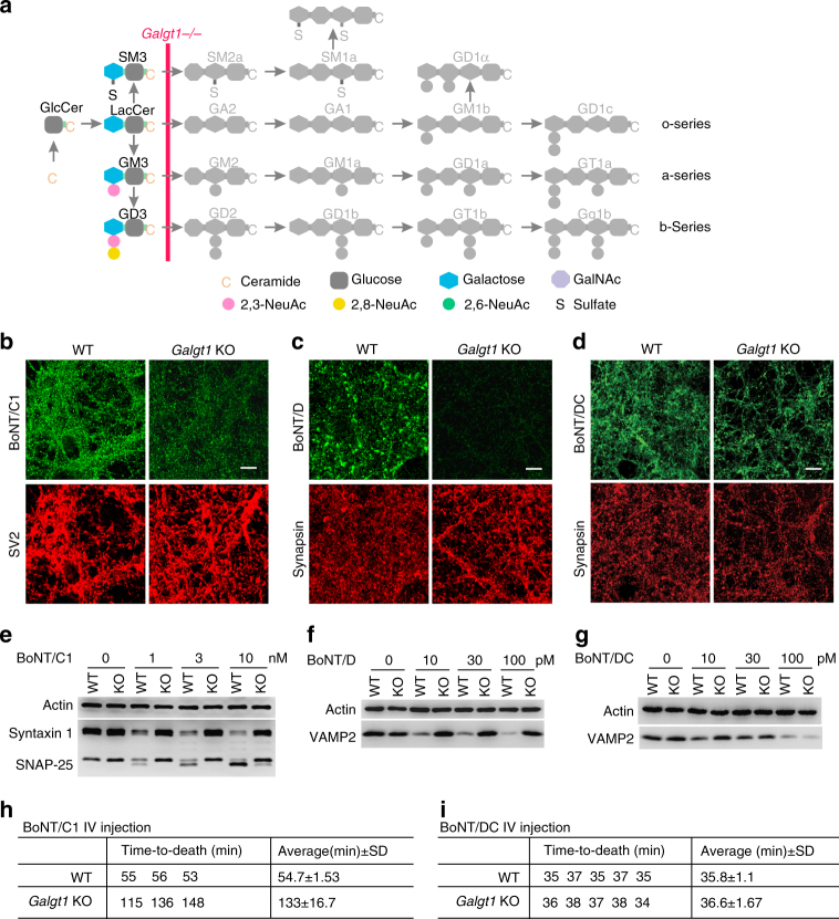
Fig. 2.
Depleting all complex gangliosides did not affect binding and entry of BoNT/DC into neurons or its toxicity in vivo. a The step blocked in Galgt1 KO mice is marked on ganglioside synthesis pathways. b–d Experiments were carried out as described in Fig. 1b–d, except with cortical neurons cultured from Galgt1 KO mice. Binding of BoNT/C1 and BoNT/D-HC was greatly reduced in Galgt1 KO neurons. In contrast, binding of BoNT/DC-HC to Galgt1 KO and WT neurons was at similar levels. e–g Experiments were carried out as in Fig. 1e–g, except with cortical neurons cultured from Galgt1 KO mice. Entry of BoNT/C1 and BoNT/D into Galgt1 KO neurons was blocked, while entry of BoNT/DC-HC into Galgt1 KO and WT neurons was at similar levels. h Rapid time-to-death assays were carried out for BoNT/C1. The same amounts of toxins (6 × 10⁵ LD50/ml, 100 µl) were injected into WT and Galgt1 KO mice. Time-to-death of each mouse was recorded. Galgt1 KO mice survived longer (133 min) than WT mice (55 min), reflecting a reduced sensitivity to BoNT/C1. SD standard deviation. i Rapid time-to-death assays were carried out for BoNT/DC as described in h (100 µl, ~8 × 10⁵ LD50/ml). Galgt1 KO mice showed survival time similar to WT mice when injected with the same amount of BoNT/DC, suggesting that Galgt1 KO and WT mice have similar sensitivity to BoNT/DC. One of two (b–d) or three (e–g) independent experiments is shown

