Fig. 6.
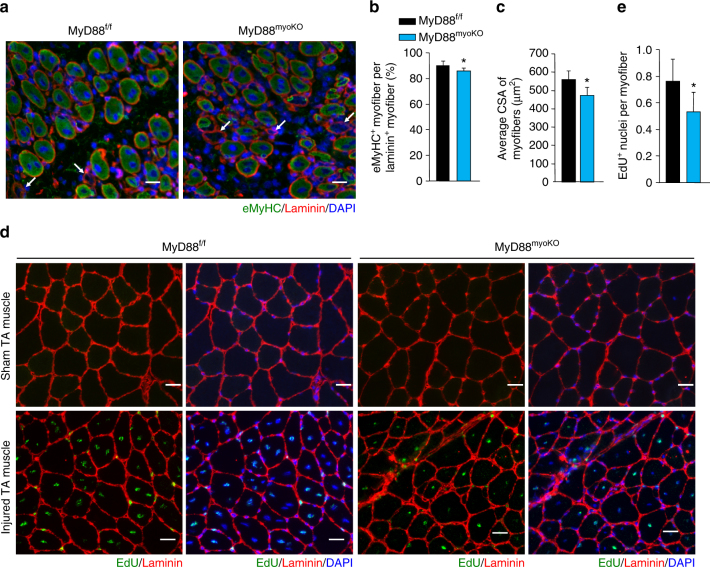
MyD88 mediates myoblast fusion during skeletal muscle regeneration. a TA muscle of 12-week-old MyD88f/f and MyD88myoKO mice was injured by intramuscular injection of 100 µl of 1.2% BaCl2 solution. After 5 days, the TA muscle was isolated and immunostained for eMyHC (green) and laminin (red) proteins. Nuclei were counterstained using DAPI (blue). Representative images are presented here. Arrows point to eMyHC−/laminin+ fibers. Scale bar: 50 µm. b Quantification of percentage of eMyHC+ myofibers within laminin staining, and c average CSA of eMyHC+ myofiber in TA muscle of MyD88f/f and MyD88myoKO mice. N = 3 in each group. *p < 0.05 from MyD88f/f mice by unpaired t-test. In another experiment, TA muscle of MyD88f/f and MyD88myoKO mice was injured by intramuscular injection of 100 µl of 1.2% BaCl2 solution. After 2 days, the mice were given an intraperitoneal injection of EdU and 11 days later TA muscles were collected and muscle sections prepared were stained to detect EdU, laminin, and nuclei. d Representative photomicrographs after EdU, laminin, and DAPI staining are presented here. Scale bar: 50 µm. e Quantification of the percentage of EdU+ nuclei per muscle fiber. N = 5 in each group. Error bars represent s.d. *p < 0.01 from MyD88f/f mice by unpaired t-test

