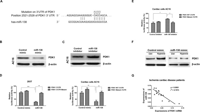
Figure 3. MiR-138 directly targets PDK1.
(A) Target prediction from TargetScan.org, microrna.org, and Exiqon. The 3′-UTR region of PDK1 contains binding sites for miR-138. (B) AC16 cells were transfected with control mimic or miR-138 mimic at 50 nM for 48 h, Western blot analysis of PDK1 protein expression was performed. (C) AC16 cells were transfected with control inhibitor or miR-138 inhibitor at 50 nM for 48 h, Western blot analysis of PDK1 protein expression was performed. A representative image is presented using β-actin as the loading reference. (D) Luciferase assay demonstrated miR-138 mimic bond to 3′-UTR of PDK1 to attenuate luciferase activity but did not affect the luciferase activity of mutant 3′-UTR of PDK1 in 293T (left) and AC16 (right) cells. (E) Luciferase assay demonstrated inhibition of miR-138 increased luciferase activity of the vector containing wild-type 3′-UTR of PDK1 but did not affect the luciferase activity of mutant 3′-UTR of PDK1 in AC16 cells. (F) AC16 cells were transfected with control mimic or miR-138 mimic for 48 h, cells were treated with or without hypoxia. Western blot analysis of PDK1 protein expression was performed. (G) Reverse correlation between miR-138 expressions and PDK1 mRNA was analyzed in cardiac tissues from ICD patients. All experiments were performed in triplicate. Data are presented with the indication of mean ± S.D. **: P<0.01; ***: P<0.001.

