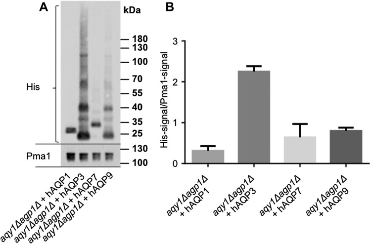
Fig. 2.
Quantification of aquaglyceroporins levels in the plasma membrane. a Quantitative Western blot analyses of aquaglyceroporins present in the plasma membrane. Protein content in plasma membrane fractions were analyzed by SDS-PAGE and Western blotting detecting Pma1 membrane marker and the polyhistidine-tag of the AQPs. Note that the typical aquaporin migrates in the gel as different mono/oligomers. b Quantification of the aquaglyceroporins present in the plasma membrane (His-signal divided by Pma1-signal), values are mean, and error bars denote SD

