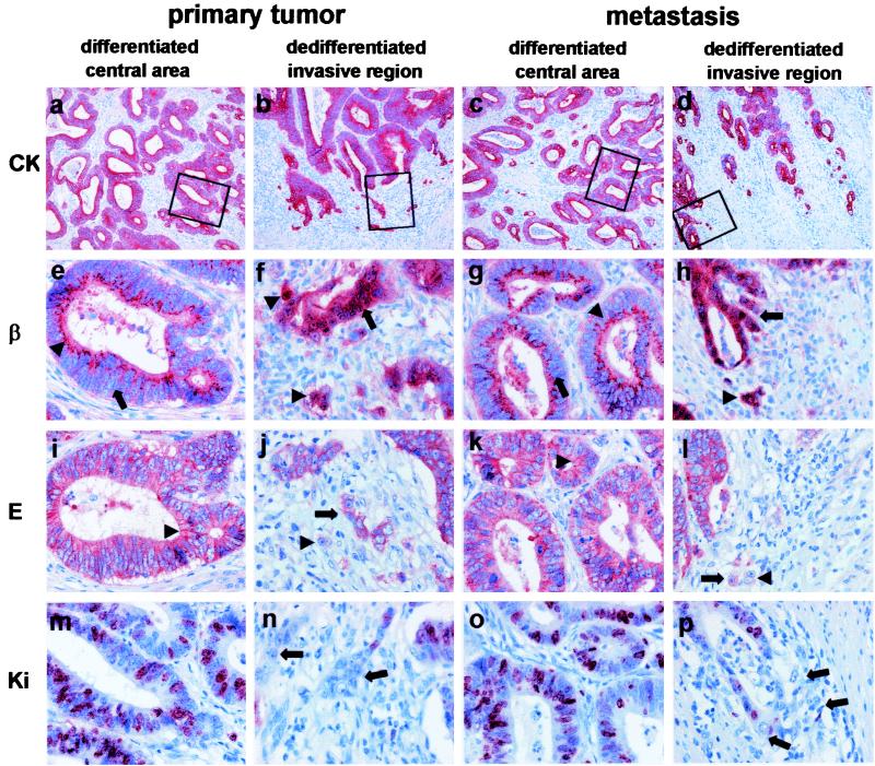
Figure 1.
Correlation of growth and the expression patterns of β-catenin, E-cadherin, and Ki-67 in well-differentiated colorectal adenocarcinomas. Shown are central areas (first column) and invasive front (second column) of the primary tumor and central areas (third column) and invasive front (fourth column) of the corresponding metastasis. Stainings are for CK-18 (first row), β-catenin (second row), E-cadherin (third row), and Ki-67 (fourth row). Boxes indicate magnified regions in stained serial sections. Specific staining is red and nuclear counterstaining is blue [×100 (a–d) and ×400 (e–p)]. CK 18 stainings show a differentiated growth pattern with tubular structures in the centers of primary tumor (a) and metastasis (c) and loss of tubular growth and tumor cell dissemination in the corresponding invasive fronts (b and d). Tumor cells are clearly polarized in the differentiated central areas in both primary tumor and metastasis, and β-catenin is localized distinctly in the apical cytoplasm and membrane of the tumor cells (arrowheads) (e and g). Note that β-catenin is not detectable in the nuclei (arrows). In contrast, tubules at the invasive front break up, and tumor cells lose their polar orientation and dissociate (arrows) (f and h). This morphological change is accompanied by nuclear accumulation of β-catenin (arrows and arrowheads). Correspondingly, tumor cells in differentiated areas of the primary tumor express membranous E-cadherin (arrowheads) (i), which is also expressed in central areas of the metastasis (k). Disseminating tumor cells at the invasive fronts with nuclear β-catenin either completely lost E-cadherin (arrowheads) or showed a cytoplasmic expression (arrows) (j and l). A high Ki-67 ratio indicating strong proliferation is found only in differentiated tubular areas of primary tumors and metastases retaining an epithelial phenotype (m and o). Disseminating, dedifferentiated tumor cells with mesenchymal phenotype did not express Ki-67 (arrows) (n and p).

