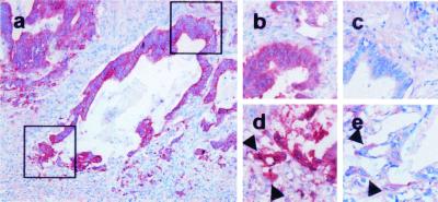
Figure 2.
Weak expression of vimentin in dedifferentiated tumor cells. CK 18 staining of a neoplastic tubulus in the invasive region of a colorectal carcinoma [×100 (a)]. Magnified region of the differentiated (upper square) or dedifferentiated area (lower square) stained in serial section against β-catenin (b and d) or vimentin (c and e). Note that dedifferentiated tumor cells with nuclear β-catenin (d, arrowheads) express vimentin weakly (e, arrowheads) but more strongly than cells in the more differentiated area (c).

