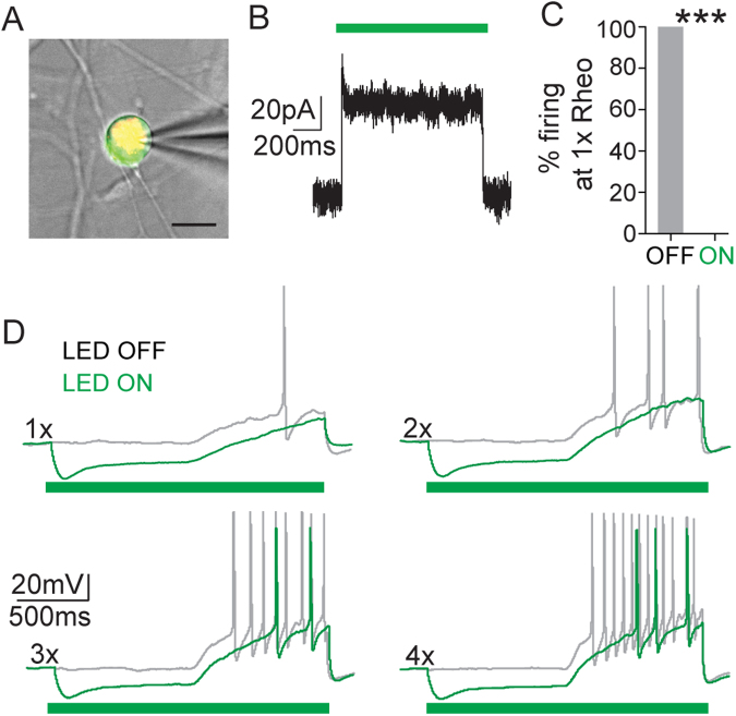
Figure 2.
Arch activation in bladder sensory neurons decreased neuronal excitability. (A) Example of a patched neuron (21 μm diameter); green fluorescence indicates Arch-GFP expression, red fluorescence is DiI labeling, indicating that the neuron projects to the bladder wall and expressed Arch (yellow); scale bar = 20 μm. (B) Representative voltage clamp recording showing photocurrent elicited by 1-second green light (530 nm) stimulation at 10 mw/mm2. (C) Quantification of the percentage of cells that showed action potential firing to a ramp current to 1x rheobase before (OFF) and during (ON) green light illumination. Optical illumination of bladder projecting SNS-Arch neurons resulted in complete blockade of action potential firing at 1x rheobase. (D) Representative traces of action potentials elicited in SNS-Arch-GFP-expressing neurons by ramp current injections of 1 to 4 times rheobase without (grey) and with (green) LED illumination. ***p = 0.001. n = 7 neurons from 4 mice; Paired t test. Error bars indicate SEM.

