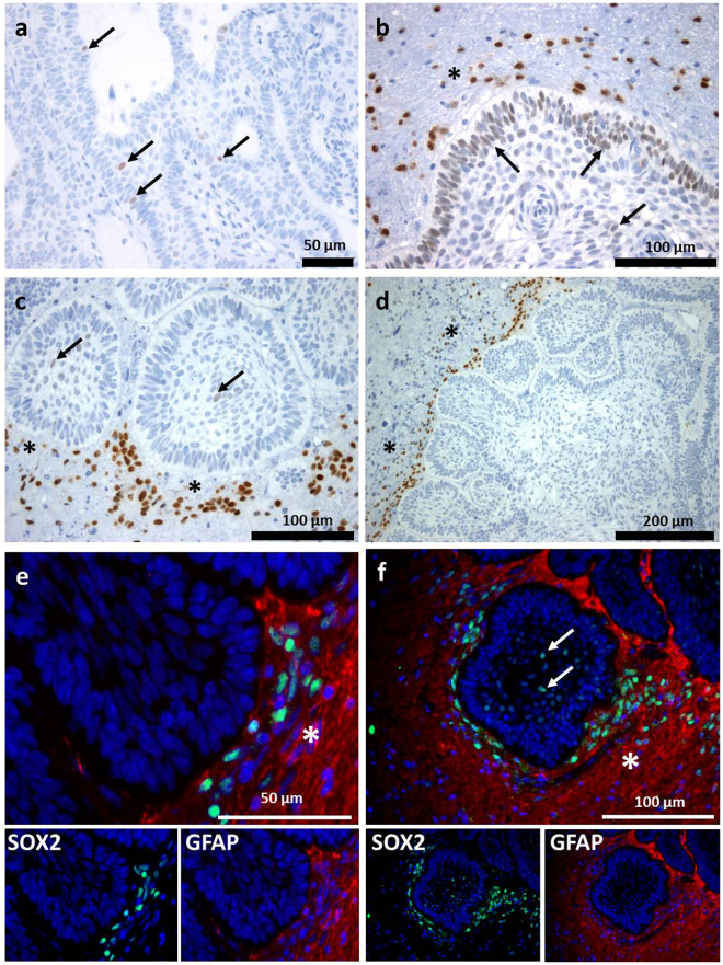
Figure 1.
Immunohistochemical Expression Pattern of Sox2 in aCP and Surrounding Brain Tissue. (a–c) Nuclear Sox2 staining (arrow) was found within aCP tumour bulk and palisading cell layer and in cells of the tumour surrounding brain tissue (asterisk; b,c,d). (e,f) Merged double-immunofluorescence staining of Sox2 (green) and the glial marker GFAP (red) demarcating adjacent brain tissue (asterisk) of the tumour revealed Sox2+ cells being enhanced nearby the CNS-tumour junction. (f) The GFAP negative tumour tissue showed also Sox2+ cells (arrow). (ada27 = a, ada56 = b, ada48 = c,d,e; ada50 = f).

