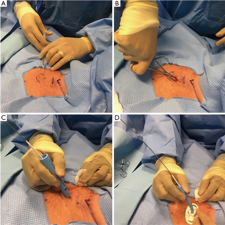
Figure 1.
PDT. (A) Infiltrating lidocaine with epinephrine in the anterior neck area. Note the skin markings on the neck, laryngeal cartilage, cricoid cartilage and the sternal notch; (B) performing blunt dissection to approach the trachea with a small Kelly clamp after incision (C) dilating the stoma with a curved tapering dilator (D) tracheostomy tube loaded on a straight dilator being inserted in the trachea.

