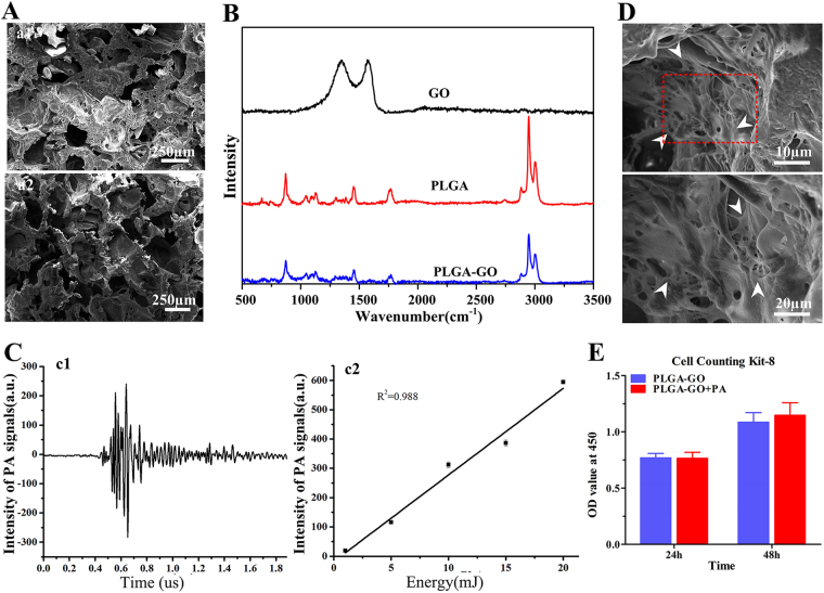
Figure 2.
Characterization of the fabricated scaffolds in vitro. (A) The microstructures of PLGA-GO scaffolds (a1) and PLGA scaffolds (a2) under the scanning electron microscope. (B) Raman spectra of GO, pristine PLGA film and PLGA-GO film. (C) Under the 10 mJ pulsed laser stimulating, graph (c1) showed the PA signal producing by PLGA-GO scaffolds; graph (c2) showed the PA signal of PLGA-GO scaffolds with different intensities of Laser. (D) Scanning electron microscopy images showed that BMSCs attached and spread well on the surface of PLGA-GO scaffold after cultivating 72 h. White arrows indicated the BMSCs. (E) Proliferation tests of BMSCs growing on PLGA-GO scaffolds with or with PA stimulation at 24 h and 48 h. All quantitative data were presented as mean ± SD, n = 4. No statistical difference was found between group as indicated by unpaired two-tailed Student’s t test.

