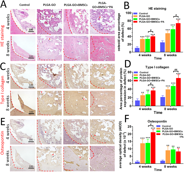
Figure 6.
Histological and immunohistochemical results indicate best outcome in animals implanting with PA-pretreated PLGA-GO combined with BMSCs. H.E staining (A) and immunohistochemical staining of type I collagen (C) and OPN (E) for analyzing the new bone formation within the sites of bone defects. The red dashed line marked the edge of the bone defect while the red arrows pointed to the GO oxide particles. Panels B, D, F are quantitative data from panels A, C, E, respectively. All quantitative data were presented as mean ± S.D, n = 5; *represents statistical difference as compared with Control group at the same time point; #represents statistical difference as compared with PA group at the same time point. *P < 0.05, **P < 0.01, ***P < 0.001; # P < 0.05, ## P < 0.01 from One-way ANOVA with Student–Newman-Keuls post hoc test.

