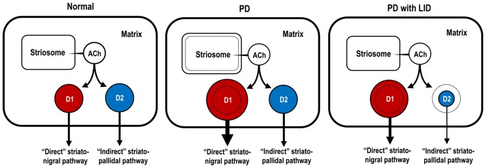
Figure 4.
Hypothetical diagram for dopaminergic regulation of Gαolf protein levels in striatonigral and striatopallidal MSNs. The sizes of the circles, colored in red and blue, indicate the abundance of Gαolf proteins in striatonigral MSNs expressing dopamine D1 receptors (D1Rs; D1-cells; red) and in striatopallidal MSNs expressing dopamine D2 receptors (D2Rs; D2-cells; blue), respectively. In the conditions of Parkinson’s disease (PD), D1-cells, but not D2-cells, might exhibit a DA D1 hypersensitivity caused by a dramatic increase in their Gαolf levels. In the conditions of PD with levodopa-induced dyskinesia (LID), D1-cells might show an increase in their Gαolf levels, while D2-cells might show a decrease in their Gαolf levels, which might result in an enhanced responsiveness to D2R activation. ACh, acetylcholine; D1-cell, striatonigral medium spiny neuron expressing DA D1 receptor; D2-cell, striatopallidal medium spiny neuron expressing DA D2 receptor; PD, Parkinson’s disease; PD with LID, Parkinson’s disease with levodopa-induced dyskinesia.

