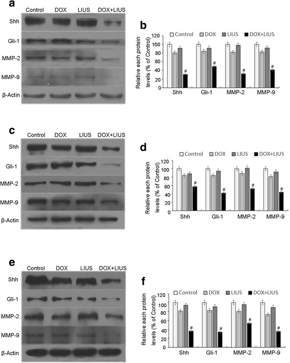
Fig. 6.

Expression of Shh, Gli-1, MMP-2 and MMP-9. Expression levels of Shh, Gli-1, MMP-2 and MMP-9 proteins were assessed by Western blotting after treatment of SAS cells (a, b), HSC-4 cells (c, d) or HSC-3 cells (e, f). β-Actin was assessed as an internal control. Representative data from three independent experiments are shown. # p < 0.05 versus control
