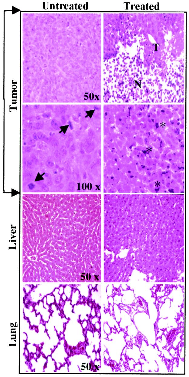
Figure 4.
Histology of tissues from untreated and Go6976-treated tumor-bearing animals. Tumor growth and treatment conditions are the same as described for Fig. 1. Tumor, liver, and lung tissues from untreated and treated tumor bearing animals were dissected 11 days after the initiation of treatment and 32 days after implantation of the cells. Tissues were processed for hematoxylin/eosin (H&E) staining, examined under a light microscope, and photographed at the indicated magnifications. Arrows show mitotic cells in untreated tumor tissue. Residual tumor (T) and necrotic cells (N) of the treated tumor are shown. Stars in the treated block indicate pycnotic cells with apparent fragmentation and clumping of nuclear DNA. Treated and untreated liver and lung tissues did not show significant microscopically detectable damages and were not different from liver and lung tissues of normal mice without tumors (not shown).

