Figure 1.

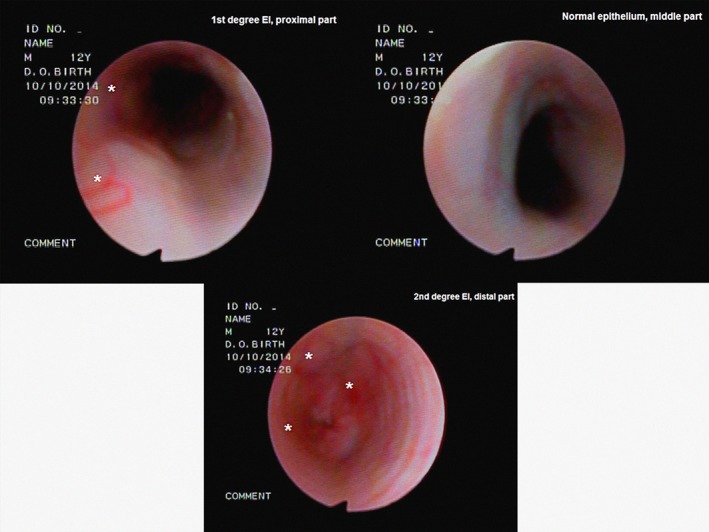
Endoscopic appearance of esophagitis:1st, normal and 2nd degree in the proximal, middle and distal part, respectively (same animal). Pathological sites are noted with an asterisk (*).

Endoscopic appearance of esophagitis:1st, normal and 2nd degree in the proximal, middle and distal part, respectively (same animal). Pathological sites are noted with an asterisk (*).