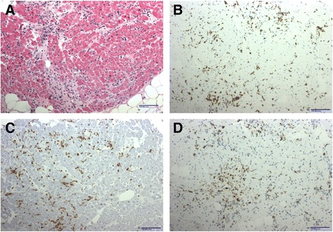
Fig. 2.

Endomyocardial biopsy revealed. a Focal mononuclear inflammatory infiltrate in early collagenized areas. b CD3 immunohistochemistry demonstrated abundant T lymphocytes while CD20 (not shown) showed only rare B lymphocytes. c CD8 immunohistochemistry showed most T lymphocytes were cytotoxic cells while CD4 staining (not shown) showed positivity in a minority of cells. d CD68 revealed also a significant number of macrophages
