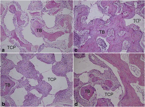
Fig. 5.

Histological evaluation of regenerated bone of PRP group constructs (a, c) and non-PRP group constructs (b, d) at 4 weeks (a, b) and 8 weeks (c, d) postoperatively. The PRP group demonstrated more extensive bone formation with the degradation of implanted constructs than the non-PRP group at each time point. (HE staining × 100. TCP, tricalcium phosphate; TB, tissue-engineered bone)
