
Figure 6. Aurora A phosphorylates CHIP at S273.
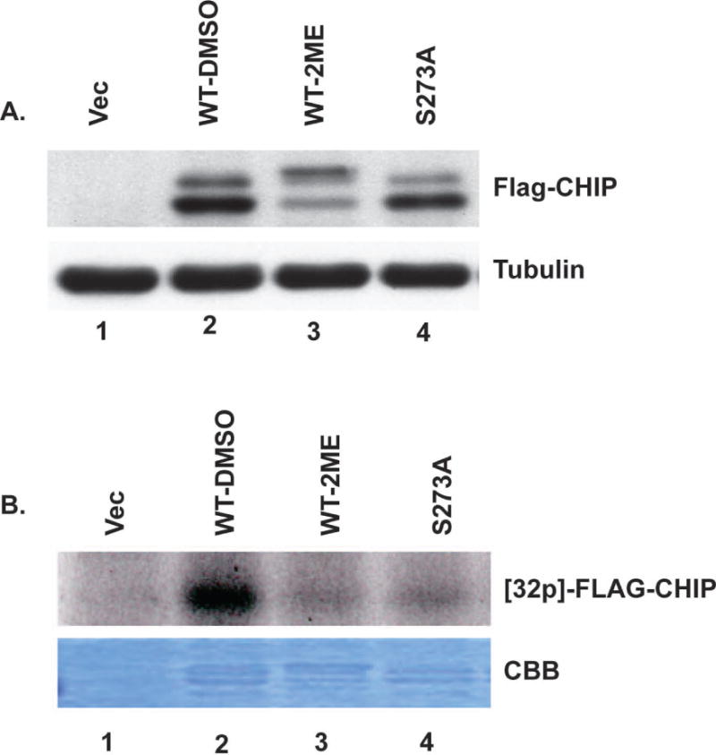
(A) Cells stably expressing FLAG-WT-CHIP were treated with or without 2-ME for 24 hrs, harvested and immunoblotted using anti-FLAG antibody (upper panel). Immunoblotting for tubulin serves as loading control (lower panel). (B) FLAG-WT-CHIP and FLAG-CHIP S273A were pulled down with M2 beads and used as substrates in kinase assays performed using bacterially expressed recombinant purified Aurora A and radiolabeled γ-ATP. Upper panel shows autoradiogram of 32P incorporated into FLAG-CHIP. Coomassie Brilliant Blue (CBB) staining of gel shows protein bands.