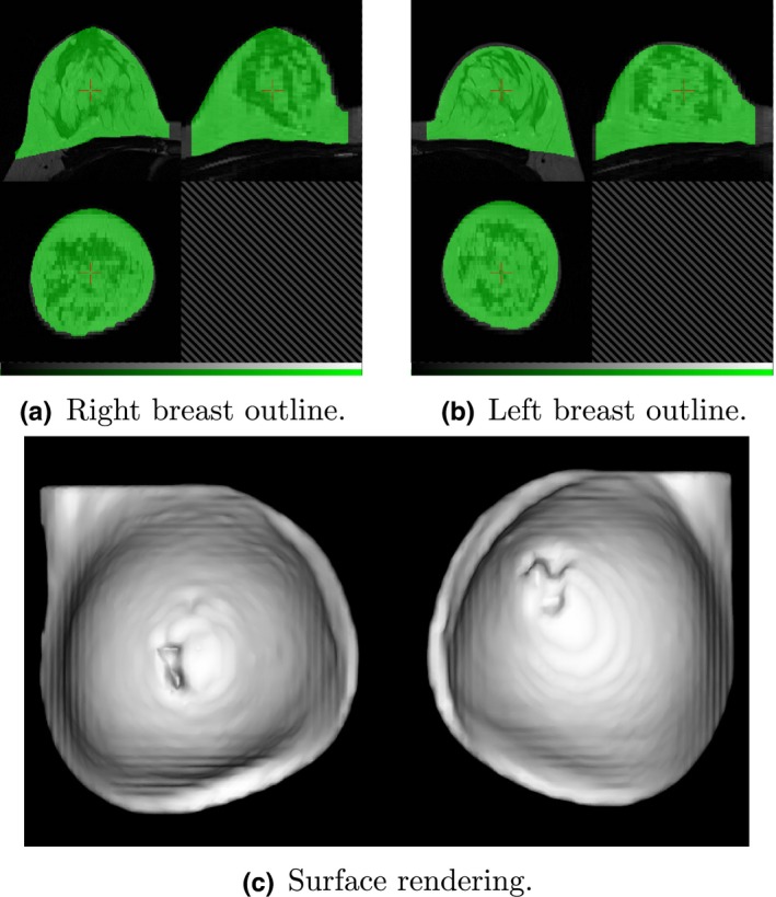
Figure 4.

Breast region mask created by removing the pectoral surface mask (Fig. 3) from the foreground mask (Fig. 2). Two views of the mask are shown, superimposed on the original MR image and centered on the right (a) and left (b) breasts. The surface rendering (c) illustrates the “squaring off” to include the axilla. [Color figure can be viewed at wileyonlinelibrary.com]
