
Figure 4.
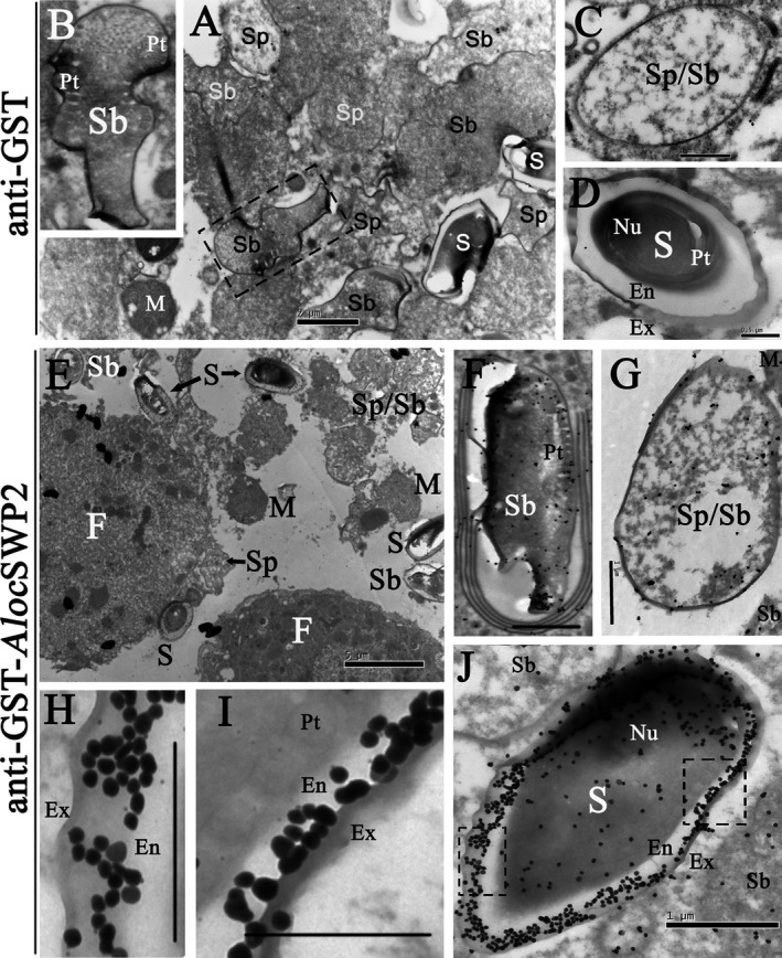
Immunocytochemistry localization of Aloc SWP2 in fat bodies of locusts. (A–D) Negative control, A. locustae‐infected locust fat cells treated with anti‐GST antibody. (E–K) Sections of A. locustae‐infected locust fat cells incubated with anti‐GST‐ Aloc SWP2 antibody. Ex = exospore; En = endospore; Pt = polar tube; Nu = nucleus; Sp = sporont; Sb = sporoblast; M = meront; S = mature spores; F = locust fat cell. Bars in D, H, I, 0.5 μm; Bars in A, C, F, G, J, 1 μm; Bar in B, 2 μm; Bar in E, 5 μm.