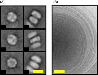
Figure 1.

Electron micrographs of: A) 1,2‐dimyristoyl‐sn‐glycero‐3‐phosphocholine (DMPC) MSP1ΔH5 nanodiscs (scale bar: 10 nm) and B) multilamellar DMPC liposomes (scale bar: 50 nm). A) The left column shows top views of the nanodiscs, whereas the right column shows side views of nanodiscs that are stacked on top of each other. Stacking arises from EM grid preparation and is a common feature of discoidal nanodiscs.5, 18
