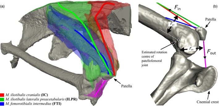
Figure 2.

(a) Anatomy of knee extensor muscles attaching to the patella in Numida. Solid lines represent lines of action for each muscle, estimated as the centre of cross‐sections taken from anatomical origin to insertion. The M. iliotibialis cranialis is shown in red, M. iliotibialis lateralis preacetabularis in green and M. femorotibialis intermedius in blue. (b) Schematic of input data for Equation (1): r ET and r PT are the moment arms of the knee extensor tendon(s) and patellar tendon, respectively, about the estimated rotation centre of the patella as it orbits the knee. Fin and Fout are the ‘input’ and ‘output’ tensions in the knee extensor tendon(s) and the patellar tendon, respectively.
