
Figure 5.
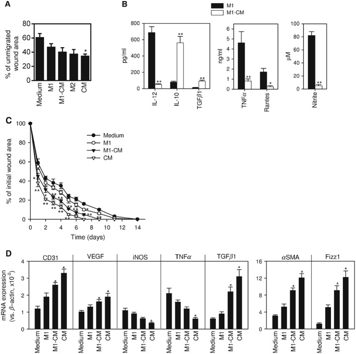
Effect of macrophage subsets in wound healing. (A) In vitro scratch wound‐healing assay was conducted with human fibroblasts plated in complete RPMI medium, used as control (medium), or with human monocytes differentiated under M1 conditions in the absence (M1) or presence of CM (M1‐CM), or under M2 conditions (M2), or with CM. The amount of wound was measured immediately after scratching (time 0) and 22 h after fibroblasts were scratched; the results are shown as mean ± SE of the percentage of remaining area of each condition, from five independent experiments; *p < 0.05 vs control medium‐treated fibroblasts. (B) Murine bone marrow‐derived macrophages were activated in the absence (M1) or presence (M1‐CM) of CM from hAMTCs; culture supernatants were assayed for cytokine levels by ELISA and for the content of nitrite (as an indirect measurement of the iNOS activity); the results are shown as mean ± SEM of 12 cultures; *p < 0.05, **p < 0.01 vs M1. (C) Murine macrophages (M1 or M1‐CM) or CM were injected into diabetic mice subjected to surgical skin wounding on the back: injection of complete DMEM was used as a control (Medium); the results show time course of wound closure after surgery and are expressed as a percentage of the original wound area; mean ± SE of eight mice/group, performed in two independent experiments; *p < 0.05, **p < 0.01 vs controls treated with DMEM. (D) Gene expression of inflammatory, angiogenic and macrophage markers in wound granulation tissue isolated 6 days after surgery; data are expressed as mean ± SE of four mice/group; *p < 0.05 vs controls treated with DMEM