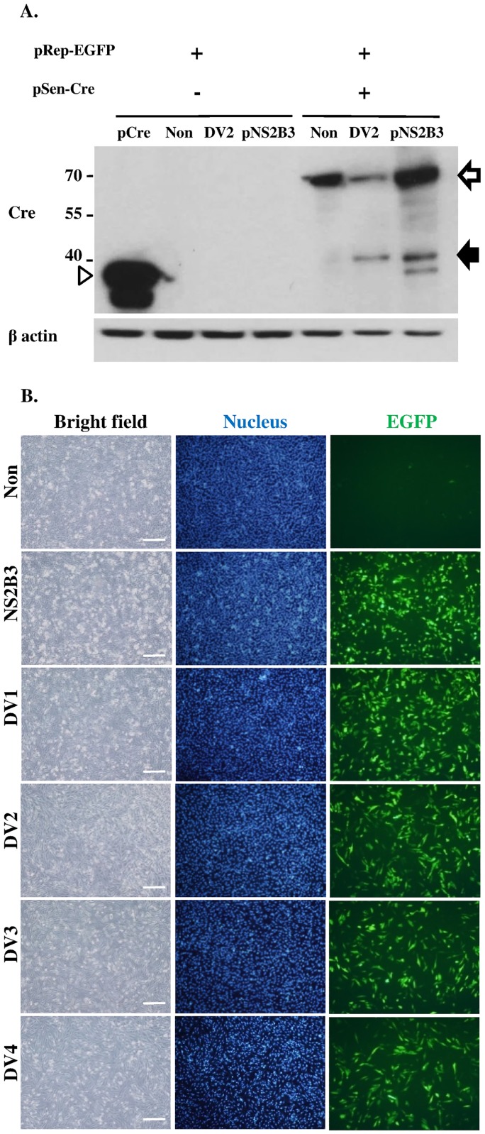
Fig 2. Characterization of DENPADS.
(A) The pRep-EGFP stable clone or F-DENPADS cells were transfected with pNS2B3 or infected with DENV-2 (m.o.i. = 5). Cells transfected with pCre or mock infected cells served as controls. Cell lysates were harvested and then analyzed by immunoblotting using anti-Cre and anti-β-actin antibodies 48 hours after transfection or infection. The cleavage product (solid arrow) represents the fragment cleaved from the NS4B-N10NS5/NLS-Cre fusion protein (hollow arrow) by NS2B3 at the region between NS4B and NS5 during DENV infection or NS3 presentation. The hollow arrowhead represents the Cre protein (~38 kDa), which is encoded by pCre. (B) F-DENPADS cells were infected with 4 DENV serotypes at an m.o.i. of 5. Cells were fixed and analyzed 48 hours post-infection using fluorescence microscopy. Scale bar, 250 μm.

