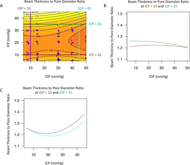
Fig 7. Change in lamina cribrosa (LC) beam thickness to pore diameter ratio with intraocular (IOP) and intracranial (ICP) pressure.
(A) Contour plot showing change in beam pore ratio as a function of IOP and ICP. Black lines indicate the contour line at the same beam thickness to pore diameter ratio. Blue dots indicate actual measurements acquired in the experiments. A sample of the contour plot at a set of (B) fixed ICP (ICP = 10mmHg, brown line; ICP = 35mmHg, dark green) and (C) fixed IOP (IOP = 10mmHg, purple; IOP = 45mmHg, light blue) conditions demonstrate the complex interaction between IOP and ICP on beam pore ratio.

