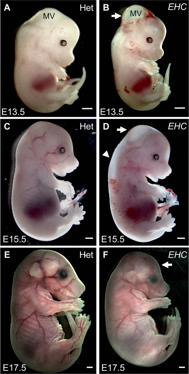
Fig 3. EHC mutant embryo morphology.
A: Heterozygous littermate at E13.5. B: EHC mutant displays hydrocephalus in the mesencephalic vesicle (white arrow). C: Heterozygous littermate at E15.5. D: EHC mutant at E15.5 displays reduced hydrocephalus (arrow), although oedema in the spinal cord region is apparent (arrowhead). E: Heterozygous littermate at E17.5. F: EHC mutant has dome-shaped head (arrow), consistent with developmental defects arising from embryonic hydrocephalus. Scale bars: 1 mm. Abbreviations: MV: mesencephalic vesicle.

