
Figure 6.
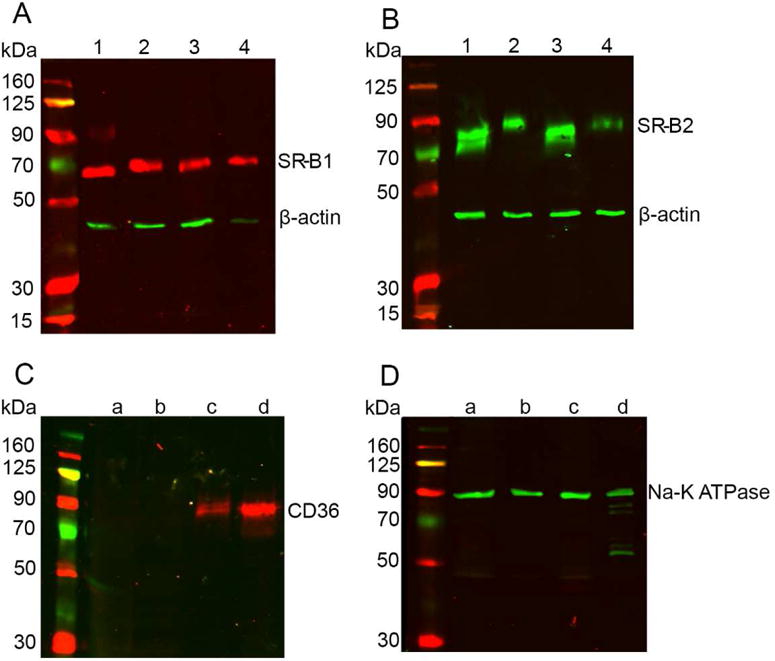
Western blots conducted on human tissues. SR-B1 and SR-B2 proteins (A and B) were detected in total tissue lysates of human macular retina (lane 1), sub-macular RPE (lane 2), peripheral retina (lane 3), and peripheral RPE (lane 4). β-actin was used as loading control. Membrane protein isolation resulted in the detection of CD36 (C) only in the peripheral RPE (lane c) and sub-macular RPE (lane d). Retinal tissue (lanes a and b) did not contain detectable CD36. Na-K ATPase was used as the loading control for membrane proteins. Panel D shows the presence of Na-K ATPase in all four tissues. Experiments were conducted in tissues from three different donors of ages ranging from 50–70 years with similar results. Results from one representative experiment are presented.