
Figure 7.
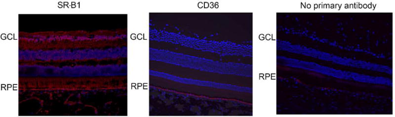
Immunohistochemistry showing expression of SR-B1 and CD36 in macaque eye sections. SR-B1 expression (red) was detected throughout the RPE and retinal layers. CD36 expression (red) was limited to the RPE layer. Both CD36 and SR-B1 antibodies were rabbit polyclonal. “No primary antibody” control sections were incubated with the secondary antibody alone. Nuclei are stained using DAPI in blue.