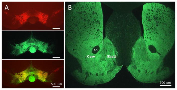
Figure 1. Expression of ChR2 in ventral tegmental area and striatum.
A. Coronal midbrain section immunolabeled to show location of tyrosine hydroxylase expressing neurons in the VTA (red, top), ChR2-eYFP expression (green, middle) and a merged overlay showing the ChR2 expression throughout the site of injection in the VTA (bottom). B. Coronal section of striatum showing expression of ChR2 in the terminal fields of midbrain dopamine neurons (A). The ventral striatum shows robust ChR2 expression in both the nucleus accumbens core and shell regions. AC; anterior commissure. Scale bar = 500 μm.

