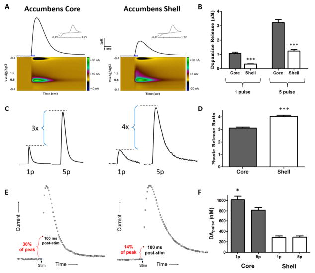
Figure 2. Light stimulated dopamine release parameters in the nucleus accumbens core and shell.
A. Representative traces of light-stimulated dopamine release in the ventral striatum. Five pulse, 20 Hz (phasic) light stimulations resulted in robust dopamine release in the nucleus accumbens core and shell. Color plots (below) display signals as current vs. voltage vs. time. Dopamine was identified by characteristic oxidation peaks at 0.6 V and reduction peaks at −0.2 V represented for both regions in the current vs. voltage plot (inset). Nucleus accumbens shell terminal fields show reduced stimulated dopamine release (peak height) and reduced dopamine uptake rates (descending portion of curve) compared to terminal fields in the nucleus accumbens core. Phasic light stimulations resulted in > 1 μM dopamine release magnitude in the accumbens shell. B. Grouped data showing dopamine release magnitude following a tonic (1 pulse) or phasic (5 pulse) light stimulation in the nucleus accumbens core and shell. Dopamine release was significantly less in the shell compared to the core for tonic and phasic stimulations. C. Representative traces show the relationship between 1p and 5p light stimulated dopamine release within nucleus accumbens core (left) and shell (right) terminal fields. D. Grouped data show phasic release as a factor of tonic release in the same field (5 pulse release / 1 pulse release). Phasic release ratios are significantly greater in the shell compared to the core. E. Representative traces show the difference in initial release velocity between the accumbens core (left) and shell (right). Dopamine measured 100 ms following the shell (14% of peak release). F. Grouped data showing that dopamine release per pulse was greater with a single pulse stimulation compared to 5 pulse stimulation in the core. Dopamine release per pulse was more uniform across stimulations in the shell. *p < 0.05, ***p < 0.001

