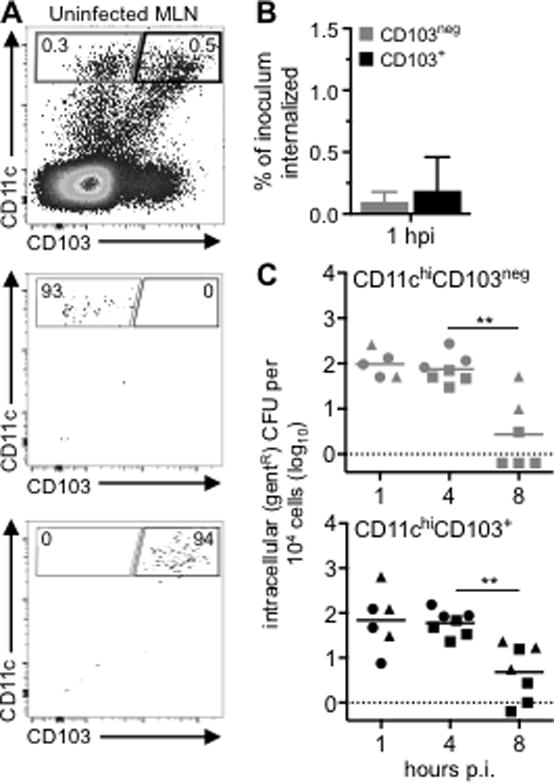
FIGURE 2.

cDC isolated from the MLN of naïve mice do not support intracellular growth of L. monocytogenes. (A) CD11chiCD103neg or CD11chiCD103+ cells were sorted from the MLN of uninfected BALB mice. Sorted cells were infected with Lm SD2000 (MOI=10–14) directly ex vivo. (B) Mean percentage (±SD) of the Lm inoculum that was resistant to 10 μg/ml gentamicin (gentR) after 1 h. (C) Intracellular growth assay for CD103neg (grey) and CD103+ (black) cDC infected directly ex vivo. Pooled data from three separate experiments (designated by circles, squares, & triangles) are shown. Cells sorted from a single mouse were used at two time points. Statistical significance was determined using Mann-Whitney analysis.
