FIGURE 5.

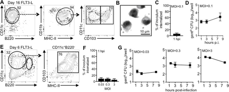
Flt3-L-cultured cells do not efficiently support the intracellular replication of L. monocytogenes. Gating scheme (A) and Diff-Quik staining (B) of bone marrow cells cultured for 16 days in 12.5% Flt3-L sup plus 0.75% GM-CSF sup. (C) Mean percentage (±SD) of Lm SD2000 inoculum that was gentR 1 h after infection of day 16 cells. (D) Intracellular growth assay using day 16 cells showing mean values (±SD) for triplicate samples. (E) Surface phenotype of bone marrow cells cultured for 6 days in 20% FLT3-L supernatant alone. (F) Mean percentage (±SD) of Lm SD2000 inoculum that was gentR 1 h after infection of day 6 cells at various MOI. (G) Intracellular growth assay using day 6 cells showing mean values (±SD) for triplicate samples. In panels C, D, F, and G data are representative of at least two separate experiments.
