
Figure 1. Sirt2 deficiency aggravates cardiac hypertrophy in aged mice.
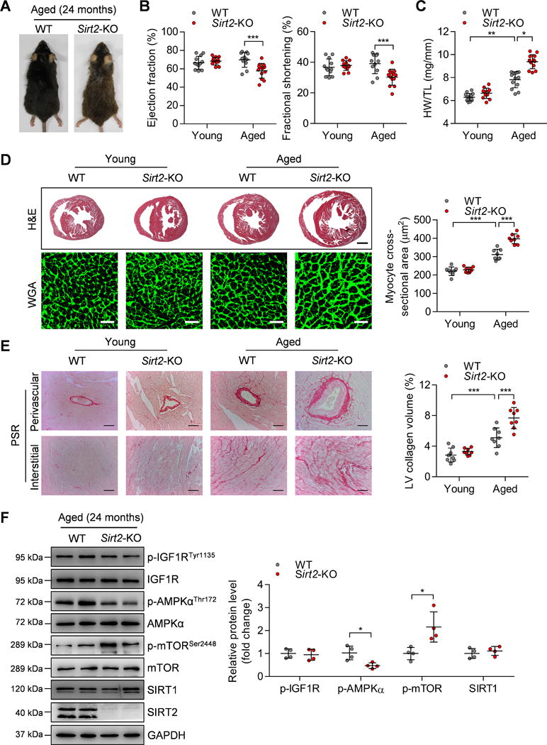
(A) Representative gross morphology of aged (24-month-old) WT and Sirt2-KO mice.
(B) Ejection fraction and fractional shortening of WT and Sirt2-KO mice at 4 months (young) or 24 months (aged). n=11~13. ***P<0.001.
(C) Heart weight-to-tibia length (HW/TL) ratios of WT and Sirt2-KO mice at 4 months (young) or 24 months (aged). n=12~13. *P<0.05, **P<0.01.
(D) Left: Hematoxylin-eosin (H&E, scale bar=1 mm) staining and wheat germ agglutinin (WGA, scale bar=30 μm) staining were performed to determine the hypertrophic growth of the hearts in young and aged WT and Sirt2-KO mice. Right: Quantification of cardiomyocyte size in young and aged WT and Sirt2-KO mice (n=8~10; ***P<0.001).
(E) Left: Picrosirius red (PSR, scale bar=50 μm) staining was performed to determine cardiac fibrosis in young and aged WT and Sirt2-KO mice. Right: Quantification of cardiac fibrosis in young and aged WT and Sirt2-KO mice (n=8~10; ***P<0.001).
(F) Left: Western blotting showing the expression levels of phosphorylated IGF1R, AMPK and mTOR and the total protein expression levels of IGF1R, AMPK, mTOR, and SIRT1. Right: Quantification of p-IGF1R, p-AMPK, p-mTOR, and SIRT1 levels (n=4; *P<0.05). IGF1R: Insulin-like growth factor 1 receptor; AMPK: AMP-activated protein kinase; mTOR: Mammalian target of rapamycin.