
Figure 2. SIRT2 expression and activity are down-regulated in hypertrophic hearts.
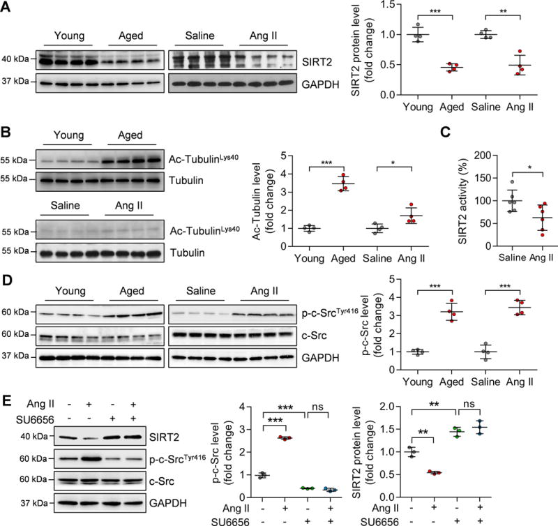
(A) Left: Representative western blotting showing changes in SIRT2 protein in aged (24-month-old) hearts and Ang II-induced hypertrophic hearts. Right: Quantification of SIRT2 protein levels (n=4; **P<0.01, ***P<0.001).
(B) Left: Representative western blotting showing changes in acetylated Tubulin levels in aged (24-month-old) hearts and Ang II-induced hypertrophic hearts. Right: Quantification of acetylated Tubulin levels (n=4; *P<0.05, ***P<0.001).
(C) SIRT2 activity in cardiac tissues of mice infused with saline or Ang II. The SIRT2 protein was purified by immunoprecipitation using an anti-SIRT2 antibody. Then an enzymatic assay was performed to determine SIRT2 protein activity (n=6; *P<0.05).
(D) Left: Representative western blotting results showing the phosphorylated level of c-Src in aged and Ang II-infused mouse hearts. Right: Quantification of phosphorylated level of c-Src (n=4; ***P<0.001).
(E) Left: Representative western blotting results showing the phosphorylated level of c-Src and level of SIRT2. Right: Quantification of phosphorylated c-Src and SIRT2 levels (**P<0.01, ***P<0.001, ns: not significant). Neonatal rat cardiomyocytes (NRCMs) were treated with Ang II (1 μM) for 24 hours with/without the presence of c-Src inhibitor SU6656 (5 μM).