
Figure 3. Sirt2-KO aggravates Ang II-induced cardiac hypertrophy.
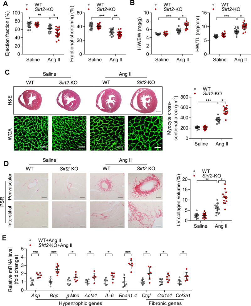
(A) Ejection fraction and fractional shortening of WT and Sirt2-KO mice treated with saline or Ang II (1.3 mg/kg/day) for four weeks (n=17~20; *P<0.05, **P<0.01, ***P<0.001).
(B) Heart weight-to-body weight (HW/BW) ratios and heart weight-to-tibia length (HW/TL) ratios of WT and Sirt2-KO mice treated with saline or Ang II (n=16~20; *P<0.05, ***P<0.001).
(C) Left: Hematoxylin-eosin (H&E, scale bar=1 mm) staining and wheat germ agglutinin (WGA, scale bar=30 μm) staining were performed to determine the hypertrophic growth of the hearts in WT and Sirt2-KO mice treated with saline or Ang II. Right: Quantification of cardiomyocyte size in WT and Sirt2-KO mice treated with saline or Ang II (n=13~17; *P<0.05, ***P<0.001).
(D) Left: Picrosirius red (PSR, scale bar=50 μm) staining was performed to determine cardiac fibrosis of the hearts from WT and Sirt2-KO mice treated with saline or Ang II. Right: Quantification of cardiac fibrosis in WT and Sirt2-KO mice treated with saline or Ang II (n=13~17; *P<0.05, **P<0.01).
(E) Quantitative real-time PCR (qRT-PCR) was performed to analyze the mRNA levels of hypertrophic (Anp, Bnp, β-Mhc, Acta1, IL-6 and Rcan1.4) and fibrosis (Ctgf, Col1a1 and Col3a1) genes (n=6; *P<0.05, ***P<0.001). Anp: Atrial natriuretic peptide; Bnp: brain natriuretic peptide; β-Mhc: Myosin heavy chain beta; Acta1: α-sarcomeric actin; IL-6: interleukin 6; Rcan1.4: regulator of calcineurin 1.4. Ctgf: Connective tissue growth factor; Col1a1: Alpha-1 type I collagen; Collagen 3a1.