
Figure 6. SIRT2 maintains AMPK signaling in myocardial tissues.
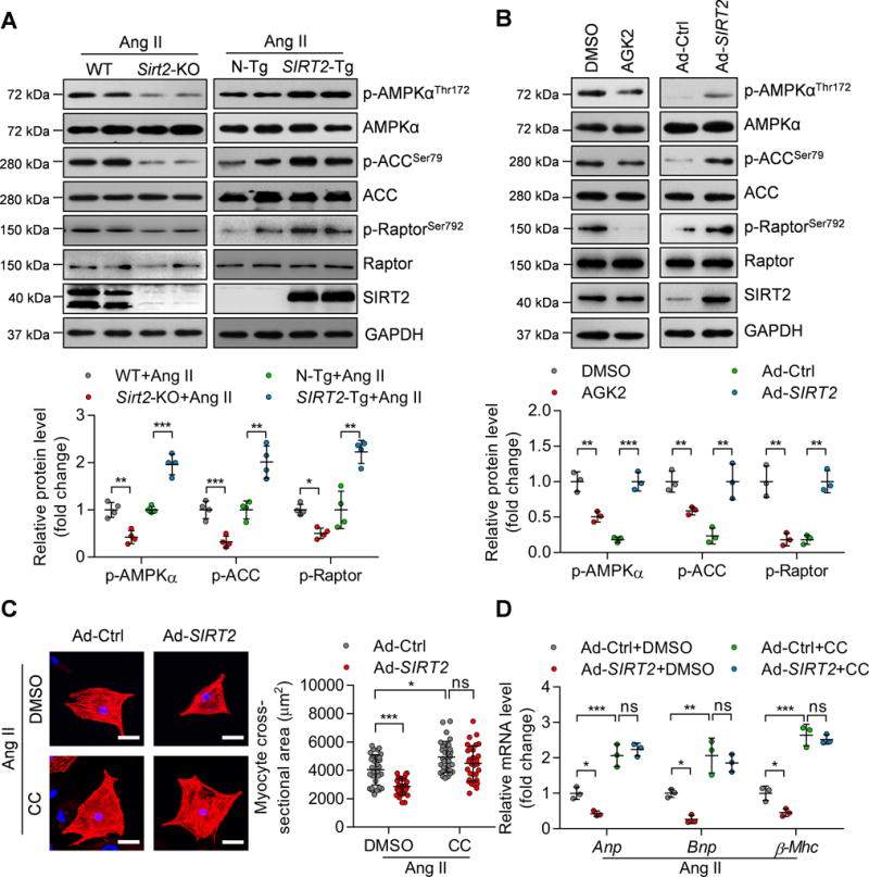
(A) Representative western blotting and quantitative results showing the phosphorylation levels of AMPK and the phosphorylation levels of its substrates ACC and Raptor in the hearts of WT, Sirt2-knockout, N-Tg and SIRT2-Tg mice infused with Ang II (n=4; *P<0.05, **P<0.01, ***P<0.001).
(B) Representative western blotting and quantification results showing the phosphorylation of AMPK, ACC, and Raptor in NRCMs. NRCMs were treated with the SIRT2 inhibitor AGK2 (10 μM) or with DMSO for 24 hours or infected with adenovirus overexpressing SIRT2 (Ad-SIRT2) or control adenovirus (Ad-Ctrl) for 24 hours (**P<0.01, ***P<0.001).
(C) NRCMs were infected with the indicated adenovirus for 24 hours and then treated with Ang II (1 μM) for 48 hours in the presence of the AMPK inhibitor compound C (CC, 10 μM) or DMSO. α-Actinin staining was performed to identify cells. Representative images (Left) and quantification of cell size of total 30 NRCMs in each group are shown (scale bar=30 μm; *P<0.05, ***P<0.001, ns: not significant).
(D) NRCMs were treated as shown in (C) and RNA was subjected to qRT-PCR to determine the mRNA level of hypertrophic genes (Anp, Bnp, and β-Mhc). *P<0.05, **P<0.01, ***P<0.001, ns: not significant.