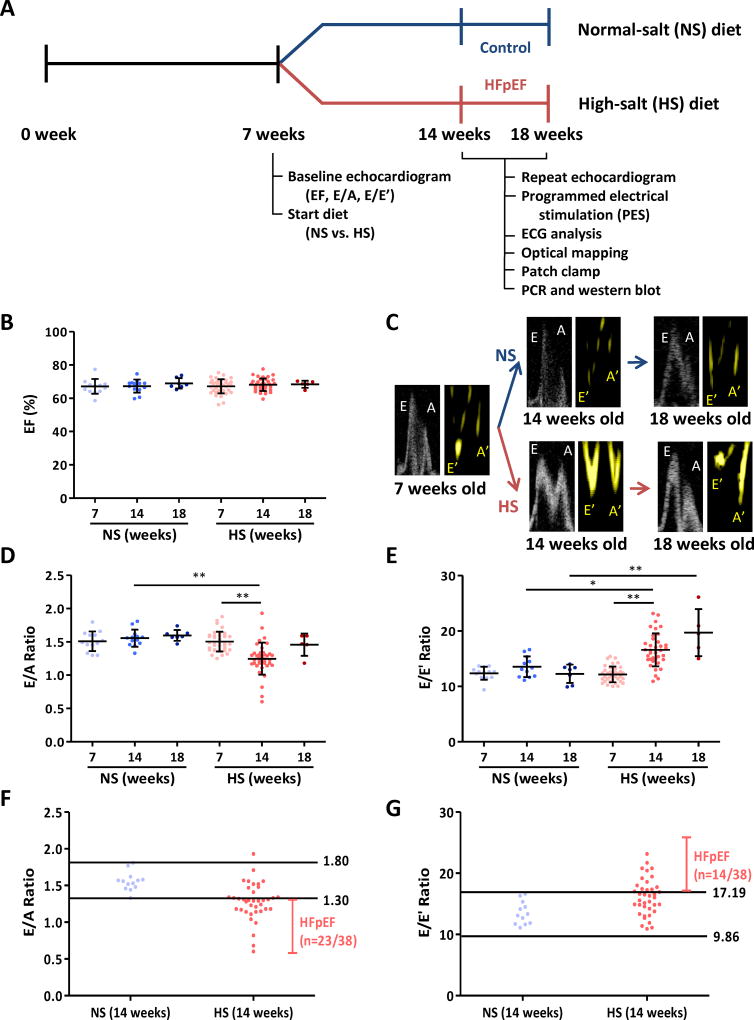
Figure 1. Timeline of experiments and echocardiographic parameters of systolic and diastolic function.
A. Dahl salt-sensitive (DSS) rats were fed high-salt (HS, n=38) and normal-salt (NS, n=13) diet at 7 weeks of age after baseline echocardiography for ejection fraction (EF) as a systolic parameter and E/A and E/E’ ratios as diastolic markers. Repeat echocardiogram was performed at 14 weeks of age. For control and HFpEF rats 14–18 weeks of age, we performed programmed electrical stimulation (PES), ECG analysis, optical mapping, patch clamp, RT-PCR and western blot. B. EF was equivalent at baseline (7 weeks) in NS and HS rats (n=13 and 38 respectively), nor did it differ in follow up in the various groups (n=13 and 38 respectively at 14 weeks, n=7 and 5 respectively at 18 weeks). C. Representative pulse wave Doppler showing E (early filling) and A (atrial filling) wave changes and tissue Doppler describing E’ and A’ wave changes in NS and HS rats at 14 and 18 weeks of age. D. Baseline E/A ratios did not differ between NS and HS rats; however, by 14 weeks E/A ratio in HS rats decreased. At 18 weeks, E/A ratio in HS rats pseudo-normalized. E. E/E’ ratio increased in HS fed rats at 14 weeks and continued to increase at 18 weeks. F. Normal value of E/A ratio was obtained from NS rats at 14 weeks old (1.30–1.80). 23 of 38 HS fed rats showed E/A ratio < 1.30, i.e. diastolic dysfunction. G. Normal value of E/E’ ratio was set (9.86–17.19) from 14-week-old NS rats. E/E’ ratio > 17.19 was shown in 14/38 HS fed rats and taken as evidence of diastolic dysfunction. NS fed rats n=13 at 7 weeks, n=13 at 14 weeks and n=7 at 18 weeks. HS fed rats n=38 at 7 weeks, n=38 at 14 weeks and n=5 at 18 weeks. Error line indicates mean and standard deviation. * denotes p < 0.05 and ** denotes p < 0.001. Mixed model regression with post-hoc testing (Tukey adjustment) was used for 1B, D and E.

