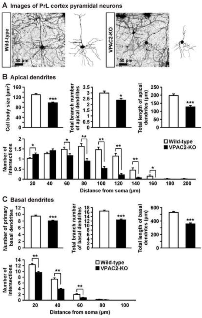
Figure 3.

Dendritic morphology of PrL pyramidal neurons in VPAC2-KO mice. (A) Golgi-stained pyramidal neuron in PrL cortex and representative tracings of the dendrites of wild-type and VPAC2-KO mice. (B, C) Cell body size and total branch number and length of apical and basal dendrites are shown. The number of intersections of dendrites with 20 μm concentric spheres centered on the soma was measured by Sholl analysis. Results are expressed as the mean ± S.E.M. of 60 neurons from 3 mice per group. *P < 0.05, **P < 0.01, ***P < 0.001, compared with wild-type.
