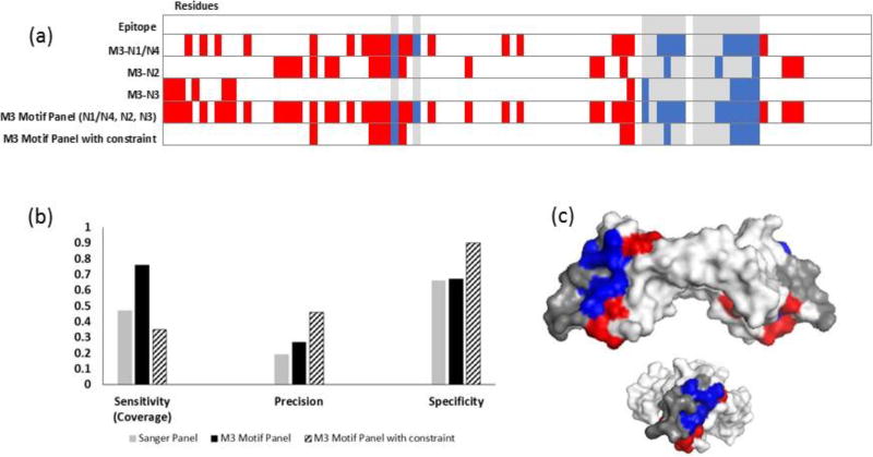
Figure 6. Increasing panel threshold improves specificity and precision, but reduces epitope coverage for bevacizumab.
(a) Heat map showing the predicted residues for each NGS motif (N1/4, N2, N3) from the final MACS round (M3), combined motif panel, and added constrain panel. (b) Adding a constraint to use only residues that were predicted by more than one motif in the panel provided improved precision and specificity but lowered epitope coverage. (c) Residues predicted with the constraint mapped onto VEGF-A (side and end views). Epitope residues colored grey were not predicted (false negatives), red residues were incorrectly identified as part of the epitope (false positives), and blue residues were correctly identified (true positives). White residues, the rest of the of antigen, are true negatives.

