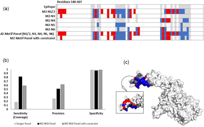
Figure 7. Increasing panel threshold improves specificity and precision, but reduces epitope coverage for trastuzumab.
(a) Heat map showing the predicted residues for each motif, combined motif panel, and added constraint panel (residues 1-540 are true negatives). (b) The compilation of NGS motifs into a panel including residues that appeared in more than one motif in the panel provided improved precision and slightly improved specificity, but lowered epitope coverage. (c) Residues predicted with the constraint mapped onto HER2 (2 sides). Epitope residues colored grey were not predicted (false negatives), red residues were incorrectly identified as part of the epitope (false positives), and blue residues were correctly identified (true positives). White residues, the rest of the of antigen, are true negatives.

