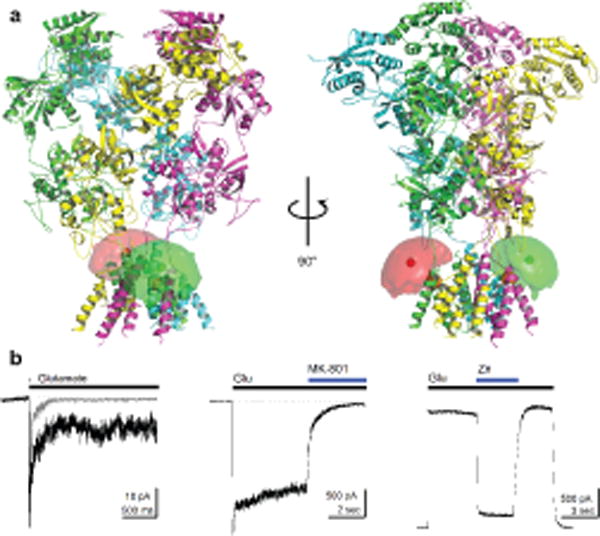
Figure 1.

smFRET constructs and characterization. (a) GluN1*F554C/GluN2A* NMDA receptors were labeled with donor and acceptor fluorophores at site 554 of GluN1, proximal to the first transmembrane segment of GluN1 (mean fluorophore positions shown as green or red hard spheres surrounded by a fluorophore cloud, and Cα of F554 on GluN1 shown as an orange sphere). (b) Representative electrophysiological responses from the smFRET construct showing deactivation (gray) and desensitization (black) (left) with 1 mM glutamate and constant 100 μM glycine recorded with outside-out patches at −60 mV, inhibition by 1 μM MK-801 recorded in whole cell mode at −60 mV (middle), and inhibition by 10 μM Zn2+ recorded in whole cell mode at +50 mV (right). Dotted lines indicate baseline current.
