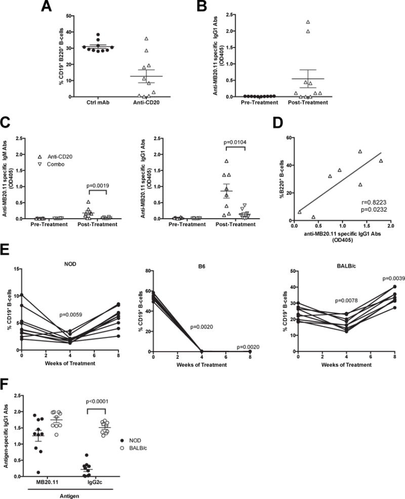
Figure 3. Differential responses to anti-CD20 (MB20.11) mediated B-lymphocyte depletion due to induction of idiotypic or isotypic directed antibodies.

A) Percentages of splenic CD19+ B220+ B-lymphocytes from NOD mice treated with anti-CD20 (MB20.11) or ctrl mAb from 10–20 weeks of age (n=10 per group). B) Sera from anti-CD20 treated NOD mice in Figure 3A were tested for the presence of anti-MB20.11 binding antibodies before and after treatment (n=10 per group). C) Sera from anti-CD20 or combo-treated NOD mice in Figure 1A were compared for the presence and isotypes (IgM or IgG1) of anti-MB20.11 binding antibodies before and after treatment (n=8 per group). D) Correlation of splenic B-lymphocytes rebounding and titer of MB20.11 binding antibodies in anti-CD20 treated NOD mice. p-value was calculated using Pearson correlation. E) Anti-CD20 mediated B-lymphocyte depletion was measured by flow cytometric analyses of CD19+ cells from peripheral blood in NOD (n=10), C57BL/6 (n=10), BALB/c (n=9) mice at 4-week-interval starting at 10 weeks of age. **p< 0.01, p-values were calculated using Wilcoxon test by comparing to pre-treatment. F) Titer of IgG1 antibodies binding anti-CD20 (MB20.11) and an irrelevant IgG2c in NOD (n=10) and BALB/c (n=9) mice after 8 weeks of anti-CD20 treatment. p-value was calculated using Mann-Whitney analysis. Each symbol represents an individual mouse; small horizontal lines indicate the Mean±SEM.
