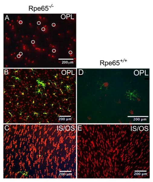
Figure 11. Mislocalized cone opsin within the OPL was used to quantify the number of stressed, surviving cones within the retina.
(A) Examples of cone opsin staining in the OPL of an Rpe65−/− retina were marked with white circles. The circles were then counted by ImageJ. (B, C) Staining of the same retinal field of a retina at different depths, OPL (B) and IS/OS (C), from a CX3CR1YFP-creERROSADTACD11cGFPRpe65−/− retina that was not TAM depleted. (D, E) Panel E shows an area of a CD11cGFPRpe65+/+ mouse retina that was well-populated with cones, and the absence of cone staining in the overlying OPL (E). This Rpe65+/+ mouse was TAM treated, but lacked the YFP-creER transgenes, so there were no YFPhi cells and no depletion by TAM; a GFPhi cell is present in the OPL (D). Panel D was enlarged to better reveal the MG-like dendritiform morphology of the GFPhi cells in this quiescent retina. Mice were sacrificed at P41 to analyze cone survival. Yellow, YFP; Green, GFP; Red, cone S-opsin.

