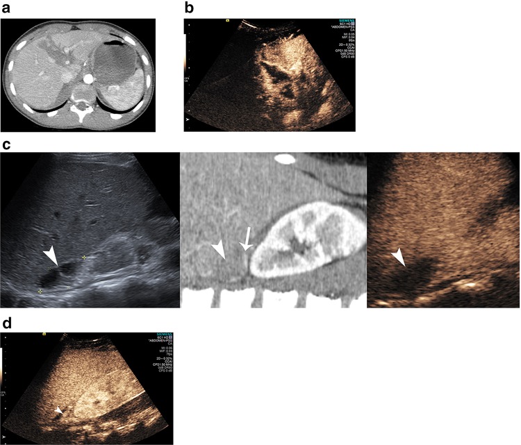
Fig. 2.
A 15-year-old boy with hepatic laceration, transection of the pancreatic neck and adrenal hematoma. Axial CT a shows the hypodense laceration affecting segments I, IVA and IVB. The corresponding CEUS image b readily demonstrates the non-enhancing laceration. A composite image c correlating US (left), CT (middle) and CEUS (right). The adrenal hematoma (arrowhead) appears anechoic on US, hypodense on CT and non-enhancing hypoechoic on CEUS. The inferiorly displaced adrenal gland can be identified on CT (arrow). A different plane on CEUS demonstrating the normal enhancing displaced adrenal gland (arrowhead)

