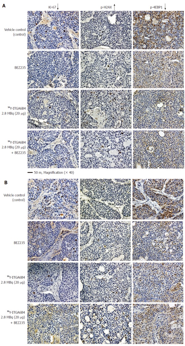
Figure 5.

Ki-67, p-H2AX, p-4EBP1 immunostaining of tumor sections. A: On Day 1 and; B: On Day 3 after administration of 90Y-ITGA6B4 alone or combined with BEZ235, intratumoral proliferation was determined by immunostaining for Ki-67 nuclear antigen. A marked reduction in Ki-67-positive cell numbers was observed in samples from mice treated with 90Y-ITGA6B4 + BEZ235 as well as with 90Y-ITGA6B4 alone and BEZ235 alone, compared with those in the untreated control sample. Meanwhile, increased p-H2AX-positive cell numbers were observed in samples from mice that received 90Y-ITGA6B4 treatment alone or the combined treatment than those in the control. Immunohistochemical analysis for p-4EBP1 showed that phosphorylation of 4EBP1 was decreased in the treatment groups compared with the control group. Tumor section images were acquired at 200× magnification and representative images are shown (scale bar, 50 μm). The quantitative and statistical analysis were summarized in Table 1.
