Fig. 1.
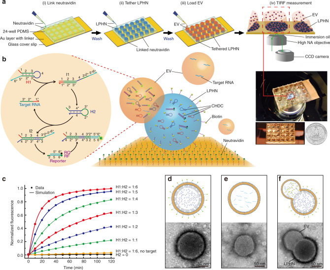
Principle and characterization of LPHN–CHDC biochip. a Stepwise operation of LPHN–TIRF assay, which is composed of link neutravidin (i), tether lipid-polymer hybrid nanoparticle (LPHN) (ii), load extracellular vesicle (EV) (iii), and TIRF measurement (iv). b Schematic illustration of catalyzed hairpin DNA circuit (CHDC) consisting of H1, H2, and reporter for signal amplification of target RNA in LPHN–EV complex (right). One target RNA catalyzes the hybridization of H1 and H2 through toehold-mediated strand displacement reactions for multiple cycles, which further destabilizes reporter moiety and generates amplified fluorescence (left). c Demonstration of catalysis. Different molar ratios of H1–H2 were introduced into CHDC at t = 0. H1 = reporter = 80 pmol. 0.0 is the background fluorescence of the absence of H2 and 1.0 is the fluorescence of H1:H2 = 1:6 at t = 120 min. The control traces (black and yellow) show the reaction with no H2 and no target GPC1-DNA, respectively. d–f Schematic drawings and transmission electron microscopy (TEM) images of LPHN (d) pancreatic cancer-derived EV (e), and LPHN–EV complex (f)

