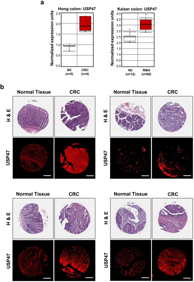
Figure 1.
Overexpression of USP47 in CRC. (a) Data obtained through Oncomine indicate higher levels of USP47 than surrounding normal tissues in two CRC subtypes. (b) Representative immunofluorescent images for USP47 protein expression in normal and CRC tissues. Samples from a human CRC tissue microarray containing colorectal carcinoma and adjacent normal tissues were examined by immunofluorescence staining with an anti-USP47 antibody. Hematoxylin and Eosin (H&E) images were provided by US Biomax Inc. Scale bar = 200 μm.

